M. Steger, S. M. Janke, P. C. Sercel, B. W. Larson, H. Lu, X. Qin, V. W. Yu, V. Blum, and J. L. Blackburn, On the optical anisotropy in 2D metal-halide perovskites, Nanoscale 14, 752‑765 (2022). doi: 10.1039/d1nr06899g.
Bis(phenylethylammonium) lead iodide: atomic structure
See all entries for this property (31 total)
Method: DFT-PBE plus Tkatchenko Scheffler dispersion, fully optimized
Origin: computational
Crystal system: triclinic
| a: | 32.35884857 Å |
| b: | 8.66248703 Å |
| c: | 8.698830605 Å |
| α: | 89.37708282° |
| β: | 85.6195755° |
| γ: | 85.42710876° |
Sample type: single crystal
- data set 225 (atomic structure)
- data set 1885 (atomic structure)
- data set 1901 (atomic structure)
- data set 1903 (atomic structure)
- data set 2267 (band gap (fundamental))
- data set 2269 (band structure)
Code: FHI-aims
Level of theory: DFT
Exchange-correlation functional: PBE+TS
K-point grid: 2*8*8
Level of relativity: atomic ZORA
Basis set definition: tight
Numerical accuracy: force convergence 5e-3 eV/AA
Comment: The original structure is based on the experimental resolved (PEA)2PbI4 structure from the reference tag, published in https://doi.org/10.1021/acs.inorgchem.7b01094
Entry added on: Feb. 25, 2020, 2:01 p.m.
Entry added by: Xixi Qin Duke University
Last updated on: March 6, 2023, 12:01 p.m.
Last updated by: Volker Blum Duke University
Download data
Bis(phenylethylammonium) lead iodide: band gap (fundamental)
See all entries for this property (3 total)
Method: Ellipsometry
Origin: experimental (T = 293.0 K)
Space group: P-1
Crystal system: triclinic
| Band gap (fundamental), eV |
|---|
- temperature = 293.0 K
Sample type: single crystal
- data set 2260 (exciton binding energy)
- data set 2261 (Exciton energy)
- data set 2266 (Exciton energy)
Starting materials: 200 mg (0.90 mmol) of PbO and 200 μL (1.59 mmol) of phenylethylammonium, fully dissolved in 4 mL of HI and 0.5 mL of H3PO2 solution.
Product: Exfoliated single crystal flakes of (PEA)2PbI4.
Description: 2D perovskite PEPI single crystals are synthesized based on previously reported slow-cooling method in Ref. https://dx.doi.org/10.1021/acsenergylett.8b01315. 200 mg (0.90 mmol) of PbO and 200 μL (1.59 mmol) of phenylethyl- ammonium are fully dissolved in 4 mL of HI and 0.5 mL of H3PO2 solution at 90 °C. The solution is then slowly cooled to room temperature at a rate of 2 °C h−1, giving orange sheet-like crystals. The crystals are then isolated from the parent solution by vacuum filtration, washed by a small amount of diethyl ether, and dried under vacuum. Thin crystals were exfoliated from the parent crystal using stiff heat release tape that serves as a handle. Sequential exfoliation steps with the tape yield successively thinner crystals. Many crystals were surveyed to select the best surface quality, flatness, and area.
Comment: Note that, while an XRD pattern was reported in this work, the XRD analysis and the space group were not reported and the space group listed here was taken from DOI: 10.1021/acs.inorgchem.7b01094.
Method: Reflection mode and transmission mode ellipsometry
Description: Transmittance was collected on a Cary 7000 UV-VIS-NIR spectrophotometer. Reflection ellipsometry was collected on a JA Woollam M2000DI at 45° to 75° using tape to suppress backside reflections. Transmission ellipsometry was collected on a JA Woollam M2000DI from −10° to 70°. The three data sets were processed as a multisample analysis in CompleteEASE. For bulk and cleaved crystals, reflection ellipsometry and reflection Mueller Matrix were collected using focus probes and either a JA Woollam M2000 or RC2, respectively.
Comment: Exciton energies were extracted from a uniaxial model of the ellipsometry data (2.385(5) eV in-plane and 2.419(7) eV out-of-plane). The exciton binding energy of 0.259 eV was calculated using ellipsometry dielectric parameters, an electron-hole image charge model and the experimental effective mass of DOI: 10.1021/acs.jpclett.0c03731.
Entry added on: March 4, 2023, 5:52 p.m.
Entry added by: Volker Blum Duke University
Last updated on: March 6, 2023, 12:01 p.m.
Last updated by: Volker Blum Duke University
Download data
Bis(phenylethylammonium) lead iodide: exciton binding energy
See all entries for this property (2 total)
Origin: experimental (T = 293.0 K)
Space group: P-1
Crystal system: triclinic
| Exciton binding energy, eV |
|---|
- temperature = 293.0 K
Sample type: single crystal
- data set 2259 (band gap (fundamental))
- data set 2261 (Exciton energy)
- data set 2266 (Exciton energy)
Starting materials: 200 mg (0.90 mmol) of PbO and 200 μL (1.59 mmol) of phenylethylammonium, fully dissolved in 4 mL of HI and 0.5 mL of H3PO2 solution.
Product: Exfoliated single crystal flakes of (PEA)2PbI4.
Description: 2D perovskite PEPI single crystals are synthesized based on previously reported slow-cooling method in Ref. https://dx.doi.org/10.1021/acsenergylett.8b01315. 200 mg (0.90 mmol) of PbO and 200 μL (1.59 mmol) of phenylethyl- ammonium are fully dissolved in 4 mL of HI and 0.5 mL of H3PO2 solution at 90 °C. The solution is then slowly cooled to room temperature at a rate of 2 °C h−1, giving orange sheet-like crystals. The crystals are then isolated from the parent solution by vacuum filtration, washed by a small amount of diethyl ether, and dried under vacuum. Thin crystals were exfoliated from the parent crystal using stiff heat release tape that serves as a handle. Sequential exfoliation steps with the tape yield successively thinner crystals. Many crystals were surveyed to select the best surface quality, flatness, and area.
Comment: Note that, while an XRD pattern was reported in this work, the XRD analysis and the space group were not reported and the space group listed here was taken from DOI: 10.1021/acs.inorgchem.7b01094.
Method: Reflection mode and transmission mode ellipsometry
Description: Transmittance was collected on a Cary 7000 UV-VIS-NIR spectrophotometer. Reflection ellipsometry was collected on a JA Woollam M2000DI at 45° to 75° using tape to suppress backside reflections. Transmission ellipsometry was collected on a JA Woollam M2000DI from −10° to 70°. The three data sets were processed as a multisample analysis in CompleteEASE. For bulk and cleaved crystals, reflection ellipsometry and reflection Mueller Matrix were collected using focus probes and either a JA Woollam M2000 or RC2, respectively.
Comment: Exciton energies were extracted from a uniaxial model of the ellipsometry data (2.385(5) eV in-plane and 2.419(7) eV out-of-plane). The exciton binding energy of 0.259 eV was calculated using ellipsometry dielectric parameters, an electron-hole image charge model and the experimental effective mass of DOI: 10.1021/acs.jpclett.0c03731.
Entry added on: March 4, 2023, 5:55 p.m.
Entry added by: Volker Blum Duke University
Last updated on: March 6, 2023, 12:02 p.m.
Last updated by: Volker Blum Duke University
Download data
Bis(phenylethylammonium) lead iodide: Exciton energy
See all entries for this property (3 total)
Origin: experimental (T = 293.0 K)
Space group: P-1
Crystal system: triclinic
| Exciton energy, eV |
|---|
- temperature = 293.0 K
Sample type: single crystal
- data set 2259 (band gap (fundamental))
- data set 2260 (exciton binding energy)
- data set 2266 (Exciton energy)
Starting materials: 200 mg (0.90 mmol) of PbO and 200 μL (1.59 mmol) of phenylethylammonium, fully dissolved in 4 mL of HI and 0.5 mL of H3PO2 solution.
Product: Exfoliated single crystal flakes of (PEA)2PbI4.
Description: 2D perovskite PEPI single crystals are synthesized based on previously reported slow-cooling method in Ref. https://dx.doi.org/10.1021/acsenergylett.8b01315. 200 mg (0.90 mmol) of PbO and 200 μL (1.59 mmol) of phenylethyl- ammonium are fully dissolved in 4 mL of HI and 0.5 mL of H3PO2 solution at 90 °C. The solution is then slowly cooled to room temperature at a rate of 2 °C h−1, giving orange sheet-like crystals. The crystals are then isolated from the parent solution by vacuum filtration, washed by a small amount of diethyl ether, and dried under vacuum. Thin crystals were exfoliated from the parent crystal using stiff heat release tape that serves as a handle. Sequential exfoliation steps with the tape yield successively thinner crystals. Many crystals were surveyed to select the best surface quality, flatness, and area.
Comment: Note that, while an XRD pattern was reported in this work, the XRD analysis and the space group were not reported and the space group listed here was taken from DOI: 10.1021/acs.inorgchem.7b01094.
Method: Reflection mode and transmission mode ellipsometry
Description: Transmittance was collected on a Cary 7000 UV-VIS-NIR spectrophotometer. Reflection ellipsometry was collected on a JA Woollam M2000DI at 45° to 75° using tape to suppress backside reflections. Transmission ellipsometry was collected on a JA Woollam M2000DI from −10° to 70°. The three data sets were processed as a multisample analysis in CompleteEASE. For bulk and cleaved crystals, reflection ellipsometry and reflection Mueller Matrix were collected using focus probes and either a JA Woollam M2000 or RC2, respectively.
Comment: Exciton energies were extracted from a uniaxial model of the ellipsometry data (2.385(5) eV in-plane and 2.419(7) eV out-of-plane). The value provide here is extracted from a cleaved crystal, in-plane.
Entry added on: March 4, 2023, 6:01 p.m.
Entry added by: Volker Blum Duke University
Last updated on: March 6, 2023, 12:02 p.m.
Last updated by: Volker Blum Duke University
Download data
Bis(phenylethylammonium) lead iodide: Exciton energy
See all entries for this property (3 total)
Origin: experimental (T = 293.0 K)
Space group: P-1
Crystal system: triclinic
| Exciton energy, eV |
|---|
- temperature = 293.0 K
Sample type: single crystal
- data set 2259 (band gap (fundamental))
- data set 2260 (exciton binding energy)
- data set 2261 (Exciton energy)
Starting materials: 200 mg (0.90 mmol) of PbO and 200 μL (1.59 mmol) of phenylethylammonium, fully dissolved in 4 mL of HI and 0.5 mL of H3PO2 solution.
Product: Exfoliated single crystal flakes of (PEA)2PbI4.
Description: 2D perovskite PEPI single crystals are synthesized based on previously reported slow-cooling method in Ref. https://dx.doi.org/10.1021/acsenergylett.8b01315. 200 mg (0.90 mmol) of PbO and 200 μL (1.59 mmol) of phenylethyl- ammonium are fully dissolved in 4 mL of HI and 0.5 mL of H3PO2 solution at 90 °C. The solution is then slowly cooled to room temperature at a rate of 2 °C h−1, giving orange sheet-like crystals. The crystals are then isolated from the parent solution by vacuum filtration, washed by a small amount of diethyl ether, and dried under vacuum. Thin crystals were exfoliated from the parent crystal using stiff heat release tape that serves as a handle. Sequential exfoliation steps with the tape yield successively thinner crystals. Many crystals were surveyed to select the best surface quality, flatness, and area.
Comment: Note that, while an XRD pattern was reported in this work, the XRD analysis and the space group were not reported and the space group listed here was taken from DOI: 10.1021/acs.inorgchem.7b01094.
Method: Reflection mode and transmission mode ellipsometry
Description: Transmittance was collected on a Cary 7000 UV-VIS-NIR spectrophotometer. Reflection ellipsometry was collected on a JA Woollam M2000DI at 45° to 75° using tape to suppress backside reflections. Transmission ellipsometry was collected on a JA Woollam M2000DI from −10° to 70°. The three data sets were processed as a multisample analysis in CompleteEASE. For bulk and cleaved crystals, reflection ellipsometry and reflection Mueller Matrix were collected using focus probes and either a JA Woollam M2000 or RC2, respectively.
Comment: Exciton energies were extracted from a uniaxial model of the ellipsometry data (2.385(5) eV in-plane and 2.419(7) eV out-of-plane). The value provide here is extracted from a cleaved crystal, out-of-plane.
Entry added on: March 5, 2023, 1:31 p.m.
Entry added by: Volker Blum Duke University
Last updated on: March 5, 2023, 1:31 p.m.
Last updated by: Volker Blum Duke University
Download data
Bis(phenylethylammonium) lead iodide: band gap (fundamental)
See all entries for this property (3 total)
Method: DFT-HSE06 (alpha=0.25, omega=(0.11 Bohr radii)^-1)+ SOC
Origin: computational
Crystal system: triclinic
| Band gap (fundamental), eV |
|---|
Sample type: single crystal
- data set 225 (atomic structure)
- data set 745 (atomic structure)
- data set 1885 (atomic structure)
- data set 1901 (atomic structure)
- data set 1903 (atomic structure)
- data set 2269 (band structure)
Code: FHI-aims
Level of theory: Spin-orbit coupled hybrid DFT
Exchange-correlation functional: HSE06 functional; exchange mixing parameter: 0.25, screening parameter: 0.11 (Bohr radii)^(-1)
K-point grid: 3x7x7
Level of relativity: Spin-orbit coupling included as follows: Self-consistent scalar relativity (atomic zero-order regular approximation) with spin-orbit coupling applied non-selfconsistently in the energy band structure calculation.
Basis set definition: All-electron; "intermediate" numerical settings and basis sets.
Numerical accuracy: Note that DFT-computed energy band gap values, even at the level of DFT-HS06+SOC, are not intended to capture the experimentally correct fundamental gap with quantitative accuracy. Rather, they are collected be comparable to other computational band gaps at the same level of theory in order to capture trends between different sources.
External repositories:
Comment: The geometry used was computationally optimized (unit cell and atomic positions) starting from the XRD-determined structure reported in https://doi.org/10.1021/acs.inorgchem.7b01094 . The level of theory used was DFT-PBE including the Tkatchenko-Scheffler van der Waals correction. The structure is available in the HybriD3 database as dataset number 745.
Entry added on: March 6, 2023, 11:45 a.m.
Entry added by: Volker Blum Duke University
Last updated on: March 6, 2023, 11:56 a.m.
Last updated by: Volker Blum Duke University
Download data
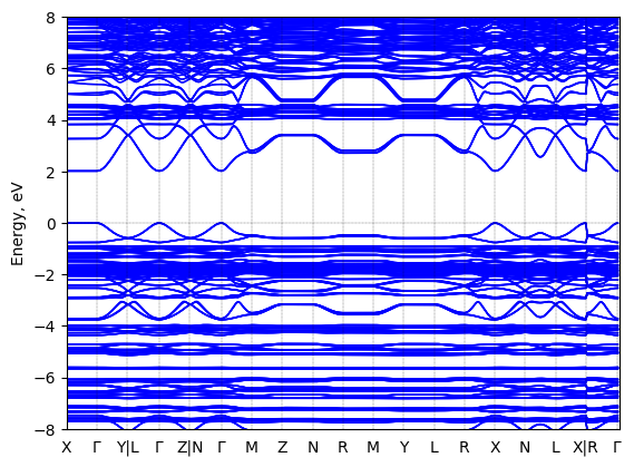
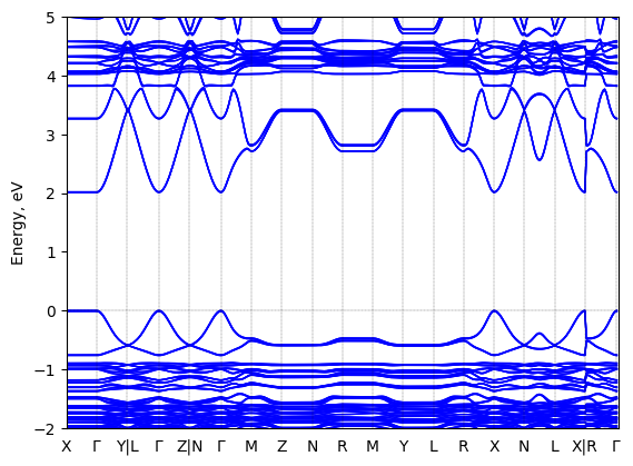
Bis(phenylethylammonium) lead iodide: band structure
Method: DFT-HSE06 (alpha=0.25, omega=(0.11 Bohr radii)^-1)+ SOC
Origin: computational
Crystal system: triclinic
Sample type: single crystal
- data set 225 (atomic structure)
- data set 745 (atomic structure)
- data set 1885 (atomic structure)
- data set 1901 (atomic structure)
- data set 1903 (atomic structure)
- data set 2267 (band gap (fundamental))
Code: FHI-aims
Level of theory: Spin-orbit coupled hybrid DFT
Exchange-correlation functional: HSE06 functional; exchange mixing parameter: 0.25, screening parameter: 0.11 (Bohr radii)^(-1)
K-point grid: 3x7x7
Level of relativity: Spin-orbit coupling included as follows: Self-consistent scalar relativity (atomic zero-order regular approximation) with spin-orbit coupling applied non-selfconsistently in the energy band structure calculation.
Basis set definition: All-electron; "intermediate" numerical settings and basis sets.
Numerical accuracy: Note that DFT-computed energy band gap values, even at the level of DFT-HS06+SOC, are not intended to capture the experimentally correct fundamental gap with quantitative accuracy. Rather, they are collected be comparable to other computational band gaps at the same level of theory in order to capture trends between different sources.
Geometry used in the calculation
Comment: The geometry used was computationally optimized (unit cell and atomic positions) starting from the XRD-determined structure reported in https://doi.org/10.1021/acs.inorgchem.7b01094 . The level of theory used was DFT-PBE including the Tkatchenko-Scheffler van der Waals correction. The structure is available in the HybriD3 database as dataset number 745.
Entry added on: March 7, 2023, 11:32 a.m.
Entry added by: Xixi Qin Duke University
Last updated on: March 7, 2023, 11:35 a.m.
Last updated by: Xixi Qin Duke University
Download data